Technology
Capture by charge, not by antibody.
Tumor cells and white blood cells can interact differently with charged surfaces. Inside a charge-tuned microtube under controlled flow, tumor cells can be retained while most white blood cells wash away. No tumor-targeting capture antibody.
Live microscopy: tumor cells retained at the wall while white blood cells move past under flow.
Results to date
What the data show so far
CTCs per 3.5 mL of patient blood
Seven patients with advanced prostate cancer. Cells were fixed, stained and classified as CTCs by an oncology lab using PSMA and cytokeratin, including EpCAM-negative CTCs.
More tumor-like events than unspiked blood
In a whole-blood spike experiment, ~100 A549 lung-cancer cells added to 7.5 mL of blood produced ~8× more tumor-like events (cytokeratin-positive, CD45-negative) than the same donor's unspiked blood processed under the same protocol.
Marker-negative CTCs recovered
EpCAM-negative CTCs were identified in blood from patients with advanced prostate cancer, alongside EpCAM-positive CTCs. Capture did not use an EpCAM-targeting antibody.
Captured cells stayed alive in culture
Published with a related version of the platform.
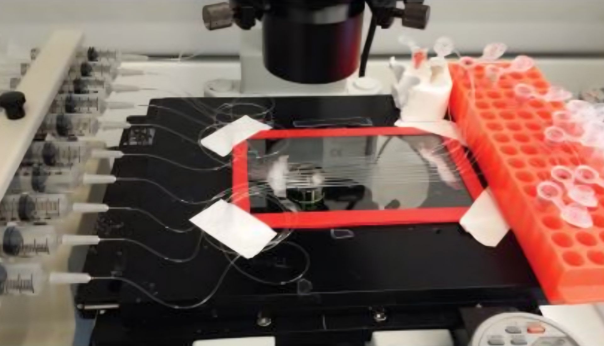
Prototype flow setup: charged microtubes on an inverted microscope.
How it works
Why charge instead of capture markers
Many tumor-cell capture methods rely on a predefined surface marker such as EpCAM. Cells with low or absent expression of that marker can be missed. Bioforce instead uses charged-surface interactions under controlled flow, so capture does not require a tumor-targeting antibody.
- The sample flows slowly through a microtube with a tuned surface.
- Tumor cells can be retained while most white blood cells pass through or wash away.
- Retained cells are released and counted.
- The lab receives the recovered cells with a QC report, for culture, analysis or other research use.
Patient evidence
Tumor cells recovered from patient blood
Blood from seven men with advanced prostate cancer, processed using the charged-surface capture approach that underlies Bioforce.
Tumor cells counted per 3.5 mL of blood
Counts vary widely between patients.
What the captured cells looked like
Stained for EpCAM, cytokeratin, PSMA, DNA (DAPI) and the white-cell marker CD45
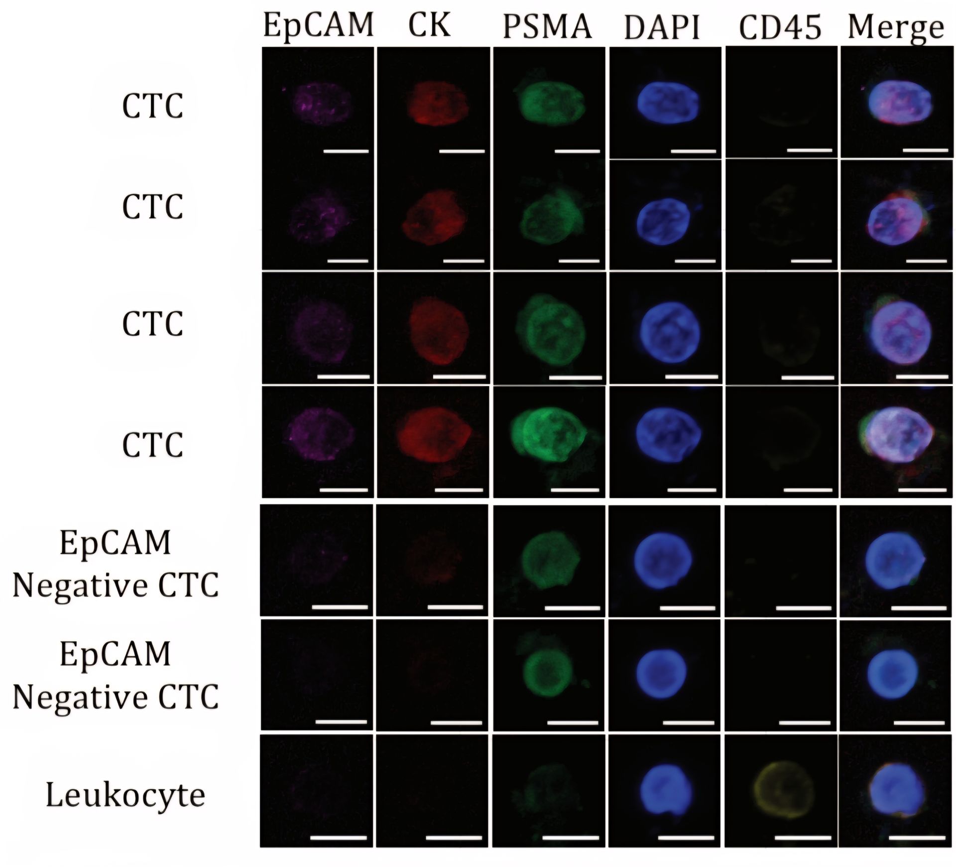
The two EpCAM-negative tumor cells (just above the white cell) are cells that EpCAM-targeted capture can miss.
Spike test
Known tumor cells, hidden in healthy blood
We added about 100 cultured lung cancer cells (A549) to 7.5 mL of healthy blood and ran it through the charge-tuned microtubes. Two 7.5 mL blood samples with no cancer cells added were processed under the same protocol. All three were stained the same way and analyzed using the same fixed computational rule.
Tumor-like events identified in each 7.5 mL sample
Same protocol and same instrument settings for all three samples
About 8 times more tumor-like events than the same donor's unspiked blood.
Reanalysis of an earlier experiment; a single spike test. A second spike with colon cancer cells, compared against 15 historical healthy-donor samples, is shown on the Data page.
Where we are
From lab method to research service
A prospective customer has sent us written requirements for the cells it needs. Our next step is meeting them from a BSL-2 lab in Phoenix: enough living tumor cells per sample, purity in whole blood, cells alive on arrival, and reliable shipping.
Need living tumor cells for your models?
Tell us what your lab needs. Pilot runs begin when our Phoenix lab opens; join the list and we will plan yours with you.
For research use only. Not for use in diagnostic procedures.